
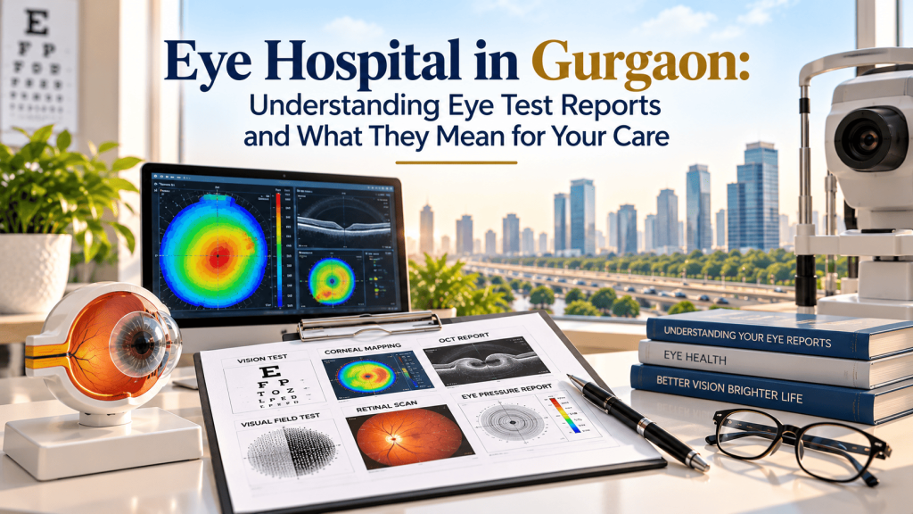
Why your eye report matters
An eye examination is more than a reading of the letters on a chart. Two people can describe the same blurred vision yet have completely different underlying problems. One may need a revised spectacle prescription. Another may need assessment of the cornea, retina, optic nerve or natural lens. Understanding what a report measures can help you ask better questions, follow your treatment plan and recognise when a change in vision needs urgent attention.
If you are looking for an eye hospital in Gurgaon, the useful question is not simply which test or machine is available. It is whether your examination answers the clinical question that brought you in. Ahooja Eye & Dental Institute (AEDI), located on New Railway Road in Gurgaon, lists services in cataract, cornea, glaucoma, retina and uvea, refractive care, contact lens care and other ophthalmic specialties. The tests you need depend on your symptoms and the ophthalmologist’s findings.
This guide explains common eye-test terms in plain English. It is educational, not a way to diagnose your own condition from a number on a report. Always have results interpreted by the clinician who examined you.
1. Visual acuity: what 6/6, 6/9 and 6/12 actually describe
Visual acuity measures how clearly you identify symbols at a set distance. In India, many reports use the Snellen notation 6/6, 6/9 or 6/12. The first number is the test distance in metres; the second corresponds to the distance at which the line is expected to be read by a person with standard acuity. A result of 6/6 means you can see that chart line at six metres. A result of 6/12 means you see at six metres what a standard observer could read at twelve metres.
This number is not a complete statement about eye health. A person can read 6/6 and still have early glaucoma, diabetic retinal changes or another disorder. Equally, reduced acuity can result from a correctable refractive error, cataract, corneal disease or a retinal problem. The difference between unaided vision and best-corrected vision gives the clinician valuable context.
Unaided vision versus corrected vision
Unaided visual acuity is measured without glasses or contact lenses. Corrected acuity is checked with the appropriate lens correction. If acuity improves considerably with glasses, a refractive error may contribute to the problem. If vision remains reduced despite refraction, the ophthalmologist may investigate other causes. The clinical significance depends on examination of the whole eye, not on one line in isolation.
2. Refraction: SPH, CYL, AXIS and near-add values
A spectacle prescription commonly includes SPH (sphere), CYL (cylinder) and AXIS. Sphere reflects the lens power used to correct short-sightedness or long-sightedness. A minus sign generally indicates myopia, while a plus sign generally indicates hyperopia. Cylinder and axis describe the amount and orientation of astigmatism. An ADD figure refers to extra near-vision power, often relevant for age-related difficulty focusing at reading distance.
A larger spectacle number does not necessarily mean a more serious eye disease. Refraction is a measure of optical correction, whereas conditions such as glaucoma or retinal disease require different assessments. Children, patients with rapid prescription changes and people considering refractive surgery may need additional checks before a final conclusion is reached.
When a prescription changes unexpectedly
A gradual adjustment can occur with age or changing refractive needs, but sudden or marked fluctuations deserve attention. Blood-glucose changes may temporarily alter vision. Cataract may also shift refraction, and corneal problems can produce irregular astigmatism. Do not treat every unexpected shift as an invitation to buy stronger glasses. Explain the timing of the change and any other symptoms to your ophthalmologist.
3. Eye pressure (IOP): a number that needs context
Intraocular pressure, often abbreviated IOP, is the pressure inside the eye and is measured in millimetres of mercury (mmHg). Tonometry measures this pressure. Eye pressure is important in the assessment and ongoing management of glaucoma, but no single reading can confirm or exclude the disease.
Some individuals develop glaucoma despite pressure readings in the statistical normal range. Others have elevated eye pressure without established optic-nerve damage. Corneal thickness, measurement technique, optic-nerve appearance, visual fields and past readings can all influence clinical interpretation. Your clinician may repeat the test or recommend further evaluation rather than diagnosing glaucoma from one number.
AEDI’s glaucoma service describes tonometry, gonioscopy, visual-field analysis and optical coherence tomography among its assessment methods. Learn about the service: https://aedi.in/best-eye-hospital-gurgaon/glaucoma-treatment/
4. Optic nerve assessment and OCT: looking beyond sharp central vision
The optic nerve carries visual information from the eye to the brain. During an eye examination, a specialist may inspect the optic-disc appearance and use optical coherence tomography (OCT), a non-invasive imaging technique that creates cross-sectional views of ocular structures. Depending on the scan protocol, OCT may examine the retinal nerve-fibre layer, macula or other features.
An OCT printout often shows colour-coded comparisons with a reference database. Red or yellow highlighting can indicate a measurement outside a selected reference range, but colour alone is not a diagnosis. Scan quality, anatomical variation, high myopia and clinical history can influence the result. Clinicians compare images over time when monitoring suspected or confirmed disease.
OCT of the optic nerve and OCT of the macula are not the same test
Optic-nerve imaging is commonly used when investigating glaucoma-related changes. Macular OCT looks at the central retina and may help evaluate swelling, fluid or structural abnormalities linked to retinal disease. A patient with distorted straight lines may therefore need a different scan from a patient undergoing routine glaucoma monitoring.
5. Visual-field testing: checking what happens outside the centre
A visual-field test measures sensitivity across different areas of vision. During an automated examination, you look at a fixation target and respond when small lights appear. It can help detect or track characteristic areas of visual-field loss associated with glaucoma and some neurological or retinal conditions.
A visual-field printout often includes indices, plots and reliability indicators. Fatigue, moving your eyes, blinking or missing instructions can affect the result. Doctors may repeat the test to establish whether a suspicious defect is reproducible. Looking at a single printout without its reliability information and clinical context can be misleading.
A key practical point: glaucoma can progress without obvious symptoms, particularly early in the disease. A person who feels they see well may still require scheduled review if their clinician identifies a risk factor or suspicious finding.
6. Corneal measurements: topography, tomography and thickness
The cornea is the clear front surface of the eye. Its curvature and thickness matter for focusing, contact lens fitting and assessment before some refractive procedures. Corneal topography maps surface curvature, while tomography can provide information about front and back corneal surfaces and thickness distribution. Pachymetry measures corneal thickness.
These investigations can be relevant when a cornea specialist in Gurgaon assesses irregular astigmatism, suspected keratoconus or suitability for laser vision correction. They may also be useful before some cataract procedures when corneal health or astigmatism is an important consideration. An isolated measurement cannot settle whether someone is suitable for surgery.
AEDI’s cornea service describes assessment and management of keratoconus, corneal infections, scars, dystrophies and other anterior-segment problems. Service information: https://aedi.in/best-eye-hospital-gurgaon/cornea-treatment/
What if the cornea is too thin for LASIK?
Corneal thickness is one factor, not the only factor. The surgeon also considers corneal shape, prescription, stability, ocular surface health and the amount of tissue a procedure would alter. Some individuals may need another treatment approach or may be advised not to undergo elective refractive surgery. Do not assume that a particular test result automatically qualifies or disqualifies you.
7. Retinal examination: why dilating drops may be recommended
The retina is the light-sensitive tissue at the back of the eye. A dilated examination allows the ophthalmologist to see more of its surface and inspect structures that a standard external eye check cannot fully evaluate. This can be particularly important for patients with diabetes, new flashes or floaters, distorted vision, or a suspected retinal condition.
After dilation, near vision may temporarily blur and bright light may feel uncomfortable. The effect varies by drop and person. Sunglasses can help, and you should ask whether driving afterwards is safe. If a clinician suspects a retinal emergency, the need for timely examination outweighs the convenience of an ordinary appointment.
Retinal imaging may include fundus photography or OCT depending on the reason for assessment. In selected cases, other imaging or tests may be recommended. No single image replaces examination, medical history and assessment of the whole eye. AEDI lists retina and uvea services here: https://aedi.in/best-eye-hospital-gurgaon/retina-uvea-services/
8. Cataract measurements: biometry and lens selection
A cataract is clouding of the natural lens. When cataract surgery is being considered, the discussion goes beyond whether a lens looks cloudy. Clinicians assess the effect on everyday tasks, examine other ocular conditions and measure the eye to plan intraocular lens (IOL) power.
Biometry measures parameters such as eye length and corneal curvature for IOL calculations. These calculations estimate which implanted lens power may suit the eye. Measurements are important, but they are not a promise of perfect vision. Coexisting retinal disease, glaucoma, corneal irregularity, healing variation and individual visual needs can affect outcomes.
Does bladeless cataract surgery change the basic need for assessment?
Whether surgery includes femtosecond-laser-assisted steps or conventional techniques, appropriate diagnosis, biometry, surgical counselling and follow-up remain essential. Surgical methods have different practical considerations; a surgeon should explain what is medically appropriate for the individual and why. A technology label on its own is not proof of superiority for every patient.
For AEDI’s description of cataract care, see https://aedi.in/best-eye-hospital-gurgaon/cataract-surgery-gurgaon/
9. Tests before LASIK or ICL: two procedures, different questions
LASIK changes corneal curvature to correct eligible refractive errors. ICL surgery places a lens inside the eye without removing the natural crystalline lens. They are not interchangeable procedures, and neither is suitable for every patient. A detailed assessment helps determine whether surgery is appropriate at all.
Before laser vision correction, the team commonly assesses prescription stability, corneal shape, thickness, tear-film and ocular health. ICL planning involves measurements relevant to lens sizing and the front of the eye, in addition to evaluation of eye health. The surgeon also discusses risks, alternatives, recovery expectations and the possibility of still needing glasses.
A consultation should therefore be an eligibility discussion, not a predetermined commitment to surgery. More about AEDI’s ICL service: https://aedi.in/best-eye-hospital-gurgaon/icl-surgery-center-gurugram/
10. How reports guide follow-up rather than giving a one-time verdict
Many eye conditions are diagnosed and managed through change over time. A baseline scan, pressure reading or visual field gives the clinician a starting point. Subsequent tests may show whether a finding is stable, progressing or affected by treatment. The goal is to combine examination findings with symptoms and risks, not simply collect reports.
For glaucoma, the team may compare pressure measurements, optic-nerve observations and visual-field results. In retinal disease, repeat imaging can help monitor structural change. In contact lens care, corneal findings and ocular-surface comfort influence continued suitability. Following the schedule agreed with your doctor is part of care even when your vision seems unchanged.
11. What to bring to an eye hospital appointment in Gurgaon
Bring your current glasses or lens prescription, recent eye reports, a list of medicines and eye drops, and details of known health conditions such as diabetes or hypertension. If you use contact lenses, tell the clinic in advance, since some measurements may require a period without lens wear. Record which eye is affected, when symptoms began and whether they are worsening.
It also helps to write down your main question: Are you worried about night driving, blurry reading vision, repeated prescription changes, eye pressure, a relative with glaucoma, or a surgical recommendation? A focused history lets the clinical team decide which investigations are useful. If dilation is likely, arrange transport as needed.
Questions worth asking after the examination
Ask what the leading diagnosis is, what the report does and does not show, whether additional tests are required, and what changes should prompt earlier review. If treatment is proposed, ask about expected benefits, alternatives, limitations, risks and follow-up. For ongoing conditions, ask which measurements are being monitored and when the next comparison should be made.
12. Symptoms that should not wait for a routine booking
Seek urgent medical assessment for sudden vision loss, a new curtain or shadow across vision, sudden flashes with many new floaters, severe eye pain, a painful red eye accompanied by reduced vision, or a significant chemical eye injury. Sudden eye pain with nausea and blurred vision can be an emergency. Do not delay care to compare hospital websites or wait for a scheduled non-urgent consultation.
Persistent but non-sudden symptoms also deserve assessment. These include difficulty seeing despite updated glasses, recurrent headaches associated with visual tasks, progressive glare, unusual distortion or recurring eye redness. Timely evaluation matters because some eye diseases begin quietly while others need rapid treatment.
Eye care at AEDI in Gurgaon
Ahooja Eye & Dental Institute is located at 560/1, Dayanand Colony, New Railway Road, Gurgaon, Haryana, India. Its official website lists eye-care services including cataract, cornea, glaucoma, retina and uvea, laser vision correction, ICL and contact lens care. Whether a particular examination or procedure is needed is decided after individual clinical assessment.
For an appointment, the official AEDI site lists Eye Care: 8130784234 and board number: 0124-2321234. Published outpatient hours are Monday to Saturday, 8:30 AM to 5:30 PM, with Sunday closed; confirm current timing and specialist availability before travelling. Appointment page: https://aedi.in/contact-us/book-appointment/
Official website: https://aedi.in/
Frequently asked questions
1. Can a normal 6/6 eye-chart result rule out glaucoma?
No. Visual acuity mainly measures clarity of central vision. Glaucoma can affect the optic nerve and peripheral visual field without noticeably reducing chart reading in early stages. A clinician assesses risk and decides whether additional testing is necessary.
2. Why did my doctor recommend OCT when I can see clearly?
OCT provides structural information about the retina or optic nerve that an eye-chart test cannot provide. It may help assess a suspected finding, establish a baseline or monitor an existing condition.
3. Does high eye pressure always mean glaucoma?
No. Eye pressure is one part of glaucoma assessment. The clinician also considers optic-nerve findings, corneal factors, visual fields and other information. Some people develop glaucoma even without elevated readings.
4. Is the red section on an OCT report a confirmed disease?
Not necessarily. Colour coding compares certain measurements with a reference group. Scan quality, anatomy and clinical context matter, so a specialist must interpret the result.
5. Why might my visual-field test need to be repeated?
The test relies on steady fixation and reliable responses. Fatigue, learning effects or missed signals can affect the result. Repetition can help determine whether a suspected change is consistent.
6. Can corneal topography tell me whether LASIK is suitable?
It provides important information about corneal shape, but LASIK eligibility also depends on thickness, prescription stability, ocular surface, general eye health and the surgeon’s judgement.
7. Are biometry readings the same as a glasses prescription?
No. Biometry records anatomical measurements used for calculations such as intraocular lens power before cataract surgery. Refraction measures the optical correction required for clearer sight.
8. Should people with diabetes have a retinal examination even without symptoms?
Yes, retinal assessment is important because diabetic eye changes may occur before symptoms are noticed. Ask your treating doctors how often a dilated retinal examination is appropriate for your situation.
9. Can I drive after having my pupils dilated?
Dilating drops can temporarily blur vision and increase sensitivity to light. Effects vary, so ask the clinic whether you should arrange a driver or other transport.
10. What is the first step if I need an eye hospital in Gurgaon for unclear symptoms?
Arrange a clinical eye examination and explain when the symptoms began, whether one or both eyes are involved, and any existing diagnoses. For sudden vision loss, severe pain or new curtain-like shadow, seek urgent care rather than a routine appointment.
Conclusion: the right decision comes from the full picture
An eye report is useful when it answers a question about your vision and helps guide an appropriate next step. A 6/6 reading, an IOP number, an OCT colour map or a corneal scan should never be interpreted on its own. If you are considering an eye hospital in Gurgaon, use your consultation to understand what each examination measures, what the findings mean for you and whether treatment or follow-up is required.
At AEDI, patients can discuss their symptoms and available ophthalmic services with the clinical team. Your diagnosis, treatment suitability and follow-up plan should always be based on an individual assessment.
