
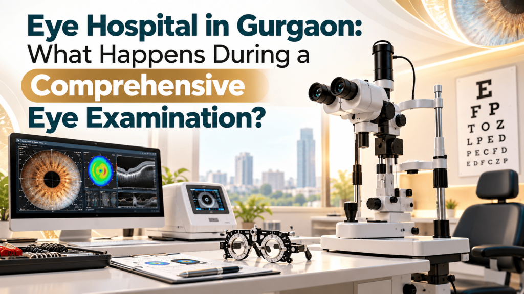
A clear vision chart is useful, but it does not tell the whole story of eye health. Someone may read small letters comfortably and still have an early eye condition that affects the optic nerve, retina or cornea. Another person may notice blurred vision and assume that the spectacle prescription has changed, when the cause is a dry ocular surface, cataract or a retinal problem. A comprehensive eye examination helps distinguish these possibilities.
For people searching for an eye hospital in Gurgaon, understanding the examination process can make the first appointment more productive. This guide explains the purpose of common tests, how doctors decide whether additional investigations are necessary, what to carry, and when symptoms require urgent care. It also explains the difference between a routine vision assessment and a medical evaluation of the eye. The exact tests are chosen by the treating ophthalmologist, not every patient needs every investigation.
What is a comprehensive eye examination?
A comprehensive medical eye examination evaluates both how well a person sees and whether the structures responsible for vision are healthy. It usually begins with symptoms and medical history, followed by vision assessment and a clinical examination. Depending on age, risk factors and findings, the ophthalmologist may examine the front surface of the eye, measure intraocular pressure, dilate the pupils to inspect the retina and optic nerve, or request focused imaging.
It is different from purchasing spectacles after a quick power check. Refraction identifies the lens prescription that can improve vision. A medical eye examination investigates why vision has changed, checks for eye disease and determines whether monitoring or treatment is appropriate. Both can be important, but they answer different questions.
Before your appointment: information that helps the eye doctor
The examination starts before any instrument is used. A useful history can direct attention to an underlying condition that might otherwise be missed. Tell the clinician when your symptoms began, whether one or both eyes are affected, and whether the change was sudden or gradual. Mention flashes, floaters, pain, redness, sensitivity to light, double vision and previous eye injuries, even if they seem unrelated to your main concern.
Bring your current spectacles, contact lens details, previous prescriptions, eye scans, surgery records and a list of medicines. In particular, disclose steroid eye drops or tablets, diabetes, high blood pressure, autoimmune disease, past laser procedures, and glaucoma in the family. If you wear contact lenses, ask the clinic whether they should be removed in advance of any planned corneal measurement. The required interval depends on the lens type and the test.
Pupil-dilating drops can blur near vision and cause light sensitivity for several hours. If dilation is likely, arrange transport rather than assuming you will be comfortable driving afterward. Allow time for the examination and discuss any accessibility, mobility or communication needs when arranging your visit.
Step 1: Visual acuity and refraction
The familiar eye chart measures visual acuity, usually with each eye tested separately and then together when relevant. Distance and near vision may be checked. Refraction determines whether myopia, hyperopia, astigmatism or age-related near-vision changes are contributing to blurred vision. The clinician may also compare how much sight improves through a pinhole or corrective lenses.
These findings are clinically useful but not a diagnosis by themselves. An eye that does not reach expected clarity with lenses may need assessment for cataract, corneal irregularity, retinal disease, amblyopia or another cause. Even a person with excellent chart vision can have certain early disorders, including glaucoma, because central acuity may remain intact until later stages.
Step 2: A careful look at the front of the eye
A slit-lamp microscope gives a magnified view of the eyelids, tear film, conjunctiva, cornea, anterior chamber, iris and lens. An ophthalmologist can look for signs of dry eye, inflammation, corneal injury, infection and lens clouding. Sometimes fluorescein dye is used to make damage to the corneal surface easier to see.
Patients who ask for a cornea specialist in Gurgaon may have symptoms such as pain, persistent redness, discomfort with contact lenses or distorted vision. A routine slit-lamp examination is often the starting point. Corneal topography or tomography can be added when irregular curvature, keratoconus or refractive surgery eligibility is under consideration. These detailed measurements are not routinely needed for every person with dry eyes.
For information about the institute’s listed cornea services, see https://aedi.in/best-eye-hospital-gurgaon/cornea-treatment/.
Step 3: Eye pressure and the optic nerve
Intraocular pressure is one part of glaucoma assessment. Tonometry measures the pressure inside the eye, but a single pressure reading cannot confirm or exclude glaucoma. Some people develop glaucomatous optic nerve damage despite pressures within a statistically usual range, while others have raised measurements without corresponding nerve injury.
The ophthalmologist may examine the optic nerve, assess the drainage angle, review corneal thickness, or recommend optical coherence tomography (OCT) and visual field testing when clinically indicated. Eye pressure also varies and can be influenced by medications, corneal characteristics and measurement methods. The value of these tests is in their interpretation together, over time where appropriate.
If you are searching for a glaucoma doctor in Gurgaon because a relative has glaucoma, bring any family details available and ask what examination interval makes sense for your risk profile. People already prescribed glaucoma drops should not stop them simply because they feel well. Many common forms of glaucoma have no obvious early symptoms.
AEDI’s dedicated glaucoma service is described at https://aedi.in/best-eye-hospital-gurgaon/glaucoma-treatment/.
Step 4: Dilated retinal examination
The retina converts light into signals sent to the brain. Dilation allows a wider view of structures at the back of the eye, including the macula, peripheral retina and optic nerve. The clinician may use a lens at the slit lamp or another instrument to examine these areas. Dilation is especially relevant when symptoms or medical history raise concern about retinal disease.
A dilated examination may identify changes associated with diabetes, age-related macular degeneration, retinal tears or other disorders. Specialised imaging such as retinal OCT, fundus photography or angiography is selected for a specific clinical question. An OCT scan is not automatically necessary for every person who attends an eye check-up, and a photograph is not a substitute for every component of a clinical examination.
Sudden new flashes or floaters, a dark curtain across vision, or sudden loss of sight need prompt assessment. Do not postpone these symptoms until a routine check-up. For information on AEDI’s listed retina and uvea services, visit https://aedi.in/best-eye-hospital-gurgaon/retina-uvea-services/.
When do additional tests become important?
An appropriate investigation answers a defined question. The most useful discussion is not “Which advanced machine will be used?” but “What are we trying to find out, and how will this result change management?” Some commonly requested tests include:
- Corneal topography or tomography, when the shape and thickness of the cornea need assessment, such as suspected keratoconus or planning for refractive correction.
- Optical coherence tomography (OCT), which can provide cross-sectional information about the retina or measurements relevant to optic nerve evaluation.
- Automated visual field assessment, which checks areas of sensitivity across the field of vision and can help monitor certain optic nerve disorders.
- Biometry and other preoperative measurements, when cataract surgery is being planned and an intraocular lens power must be calculated.
- Retinal photography or additional retinal imaging, when documentation or a more targeted evaluation is helpful.
The quality of care depends on whether the test is appropriate, whether the result is interpreted in the context of the examination, and whether there is a clear follow-up plan. A normal test does not override concerning symptoms, and an abnormal screening result does not always establish a diagnosis without further evaluation.
Why the same complaint can lead to different treatment plans
Blurred vision is a symptom, not a specific disease. Consider a patient who struggles with night driving. The possible explanation might be an outdated glasses prescription, dry eye, cataract-related glare or another visual problem. An eye examination separates these causes rather than moving directly to a procedure.
Similarly, frequent prescription changes in a teenager may call for a different evaluation from gradual near-vision difficulty in a person over 40. An older adult with diabetes may require retinal assessment even if the person reports stable vision. The right next step is guided by the history and findings, not by the popularity of a particular surgery.
Cataract, laser vision correction and ICL: why evaluation comes first
Cataract assessment
A cataract is a clouding of the natural lens. People may notice dimmer vision, glare, reduced contrast or difficulty with activities that previously felt easy. Cataract surgery is considered in relation to its effect on daily life, the overall health of the eye and an informed discussion of benefits and risks. Cataract evaluation also checks whether problems involving the cornea, retina or optic nerve might affect the expected outcome.
Some patients search for bladeless cataract surgery in Gurgaon. Femtosecond-laser-assisted techniques may assist with selected surgical steps, but they are not automatically superior for every patient or every outcome. The treating surgeon should explain which technique is appropriate, why, and what alternatives exist. See AEDI’s published description: https://aedi.in/best-eye-hospital-gurgaon/robotic-bladeless-cataract-surgery/.
LASIK and other refractive procedures
LASIK reshapes the cornea to correct selected refractive errors. Suitability requires more than knowing the spectacle number. The clinician considers prescription stability, corneal thickness and shape, dry-eye status, overall eye health, expectations and individual risk factors. Certain people should not undergo LASIK, and alternatives may include glasses, contact lenses or another procedure after specialist assessment.
Patients interested in LASIK surgery in Gurgaon should ask which tests are required, whether both eyes are suitable, how recovery and follow-up will work, and what limitations remain possible after treatment. No procedure can responsibly promise spectacle independence for every person. AEDI lists its laser vision service at https://aedi.in/best-eye-hospital-gurgaon/contoura-lasik-gurgaon/.
Implantable collamer lenses (ICL)
ICL surgery involves placing a specially designed lens inside the eye, while the natural lens remains in place. It may be considered for selected people with refractive error, including some who are not suitable candidates for corneal laser surgery. Preoperative assessment includes ocular dimensions and other safety considerations. As with any intraocular procedure, risks, alternatives, costs and the need for follow-up should be discussed individually.
Someone researching ICL surgery in Gurugram should not select between ICL and LASIK solely from an online comparison. The choice depends on examination measurements and a surgeon’s assessment.
Contact lenses: a fitting is more than selecting a power
A contact lens centre in Gurgaon may see patients who want an alternative to spectacles, people with astigmatism, and those with corneal conditions that require specialist lens designs. Safe fitting considers lens geometry, tear film, corneal condition, wearing schedule, hygiene and tolerance. Lens power alone does not establish whether a lens fits safely.
Never sleep in contact lenses unless explicitly prescribed for that use, and do not expose them to tap water. Remove lenses and seek prompt eye care if pain, worsening redness, significant light sensitivity or reduced vision develops. Contact-lens-related infections can threaten sight if treatment is delayed. Ask for a written care routine and understand when your next review is due.
Eye examination needs change with age and health
Children and teenagers
Children may not realise that what they see is different from other people’s vision. Parents may notice squinting, headaches, sitting unusually close to screens, an eye turning inward or outward, or declining school performance. A child’s assessment may include age-appropriate vision testing, alignment, focusing ability and examination of ocular health. A paediatric ophthalmologist can determine whether glasses, amblyopia management or further investigation is needed. A normal-looking eye does not guarantee normal vision.
Adults using screens for long hours
Prolonged near work can contribute to eye discomfort and reduced blinking, but it should not be used to explain every case of blur or headache. Dry-eye assessment, refraction and other tests may help identify the cause. Regular breaks, appropriate screen positioning and addressing underlying ocular-surface issues can be useful. Persistent symptoms deserve an examination rather than repeated self-treatment with over-the-counter drops.
Adults over 40
Near-vision changes become common with age, but the same stage of life also makes assessment for glaucoma and lens changes important. A person with no symptoms still may need periodic comprehensive examinations based on age and personal risk. The interval should be individualised by an ophthalmologist; someone with a family history of glaucoma may need closer surveillance than someone without known risk factors.
People living with diabetes
Diabetic retinal disease may progress before a person notices vision loss. Eye evaluation is therefore part of diabetes care, even when spectacles seem satisfactory. The schedule varies with the type of diabetes, pregnancy status and prior retinal findings. For many adults with type 2 diabetes, guidance recommends a dilated eye examination at diagnosis, with subsequent timing based on clinical advice. Good coordination between diabetes care and ophthalmology matters.
Five questions worth asking before leaving the clinic
A clear explanation is as important as a detailed examination. Before finishing your visit, ask: What is my diagnosis or the most likely cause of my symptoms? Which finding supports it? Do I need treatment now, further tests or observation? What warning signs should prompt an earlier return? When should the next review take place? These questions help patients understand what was found and what remains uncertain.
If surgery is suggested, ask for the expected benefits, reasonable alternatives, potential complications, recovery needs and what happens if treatment is deferred. Request the relevant reports and a written treatment plan. For a chronic condition, clarify who will monitor progression and whether repeat investigations are needed for comparison.
Eye symptoms that should not wait for a routine appointment
Some symptoms warrant urgent assessment, potentially through an emergency eye service. Seek immediate medical attention for sudden loss of vision, a new dark curtain or shadow, a chemical splash, penetrating injury, severe eye pain with nausea or halos, or a painful red eye associated with reduced vision. New flashes and floaters also warrant prompt ophthalmic evaluation, particularly when they are sudden or accompanied by shadows.
Do not try to neutralise a chemical splash with another chemical. Begin rinsing immediately with plenty of clean running water and arrange emergency medical care. Do not rub an injured eye or attempt to remove an embedded object. If a routine clinic cannot assess an emergency promptly, use an appropriate emergency service rather than waiting for the next available elective slot.
Understanding care at Ahooja Eye & Dental Institute in Gurgaon
Ahooja Eye & Dental Institute (AEDI) describes ophthalmic services covering cataract, cornea, laser vision correction, glaucoma, contact lenses, paediatric ophthalmology, retina and uvea, and other eye specialties. The institute also lists dental services, which are separate from the scope of this eye-examination guide. The service required for an individual patient should be confirmed at the appointment rather than inferred from a keyword or an online search result.
AEDI’s publicly listed address is 560/1, Dayanand Colony, New Railway Road, Gurgaon, Haryana, India. For an appointment, the website lists eye contact number 8130784234 and board number 0124-2321234. Patients can use the website https://aedi.in/ or the listed appointment page https://aedi.in/contact-us/book-appointment/ to check current arrangements. Service availability, consultation schedules and appointment details should be confirmed directly before travelling.
The purpose of choosing an eye hospital in Gurgaon is to receive an evaluation that matches the symptom and risk profile. A useful consultation should leave you understanding the findings, the options, what needs monitoring and when to seek further advice.
Frequently asked questions
1. Is an eye test for glasses the same as a complete eye examination?
No. A spectacle test measures refractive error, while a comprehensive examination also evaluates eye health, symptoms and risk factors. The doctor decides which additional assessments are needed.
2. Can I have an eye disease even if I see clearly?
Yes. Some conditions, including early glaucoma and certain retinal disorders, may not affect central chart vision at first. This is why risk-based medical examinations matter.
3. Will my pupils always be dilated?
Not always. Dilation is commonly used to examine the retina and optic nerve, but the decision depends on the reason for the visit and clinical findings. Ask whether driving afterward may be affected.
4. Should I bring old eye reports to a new doctor?
Yes. Prior prescriptions, visual fields, OCT scans and surgery reports can help the ophthalmologist compare changes and avoid unnecessary repetition where appropriate.
5. Does every patient need an OCT scan?
No. OCT can be useful for specific retinal and optic nerve questions, but it is not a compulsory test for every eye appointment. Its value depends on the clinical indication.
6. If my eye pressure is normal, can I still have glaucoma?
Yes. Some patients have optic nerve damage despite pressure readings in the usual range. A glaucoma evaluation considers the optic nerve and other evidence, not pressure alone.
7. Can I book cataract surgery without a full evaluation?
A preoperative assessment is required to establish the diagnosis, assess general eye health and obtain lens-related measurements. The surgeon also discusses technique, expectations and risks.
8. Is LASIK suitable for everyone who wears spectacles?
No. Suitability depends on refractive stability, corneal measurements, ocular surface health and other factors. A specialist assessment is needed before deciding on treatment.
9. How soon should sudden new floaters be checked?
Seek prompt ophthalmic assessment, especially if floaters occur with flashes, a shadow or a curtain in vision. A retinal tear or detachment must be ruled out.
10. How often should a person with diabetes have an eye examination?
The schedule depends on diabetes type, retinal findings and other circumstances. People with type 2 diabetes are generally advised to have a dilated retinal assessment at diagnosis, then follow a clinician-directed review schedule.
Conclusion
A thorough eye appointment is a process of clinical reasoning, not simply a list of machines or a decision about surgery. The history, vision tests, examination of the front and back of the eye, and any targeted imaging should fit together to explain symptoms and guide the next step. Whether the concern is cataract, glaucoma, contact lenses, retinal health or refractive surgery, informed decisions begin with an appropriate examination and an understandable care plan.
Medical note: This article provides general information and does not replace an individual ophthalmic examination. Urgent symptoms require prompt medical assessment.
